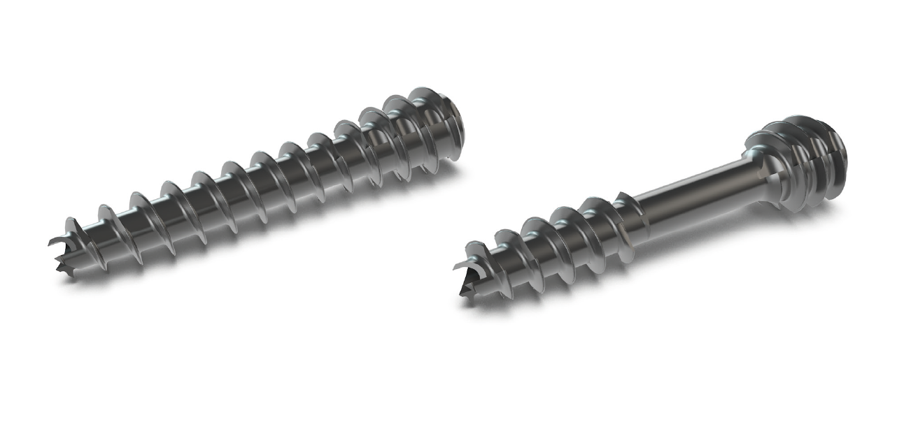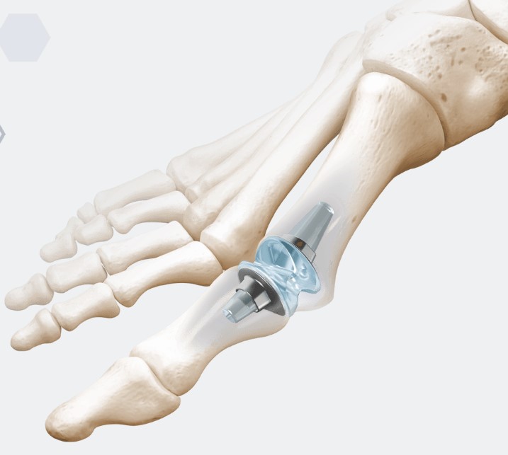
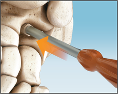
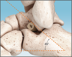
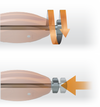
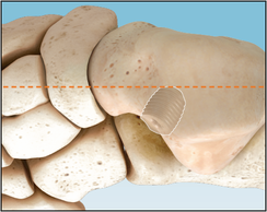
LSM – Cannulated Superscrews
Features:
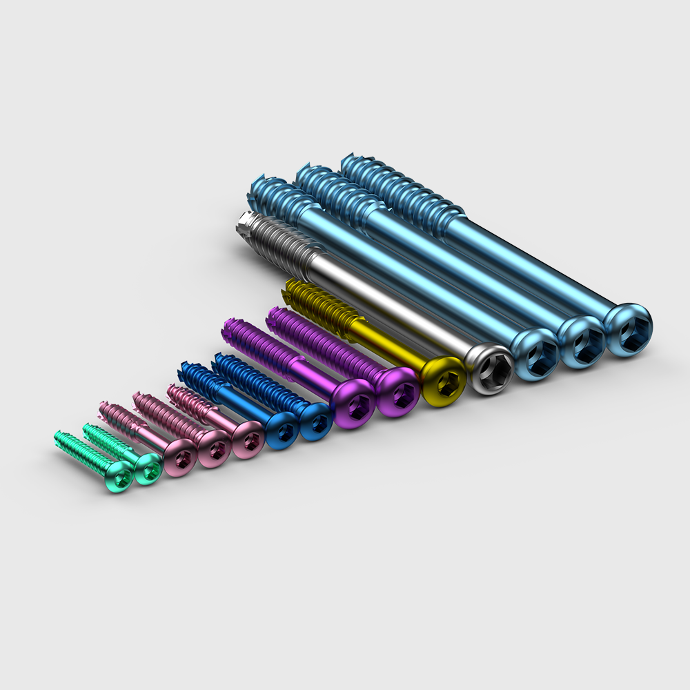
Screws with a 2.7 mm diameter are targeted at hand surgery, foot surgery, and small bone fractures.
Screws with a diameter ranging from 3.5 to 4.5 mm are aimed to tackle fractures of lateral malleolus, distal tibia (including intra-articular), radius, ulna, olecranon, distal humerus, and pelvic ring.
Screws with a diameter that ranges from 6.5 to 7.2 mm are used in femoral neck and proximal femur fractures (including basocervical and subtrochanteric), femoral condyles, tibial plateau, and ankle arthrodesis.
Discover More