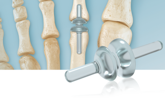
RTS Lesser Toe – MTP Joint Replacement


For Treatment of Degenerative, Painful and/or Alignment Deformities of the Lesser MTP Joints.​ Designed to Eliminate Pain and Restore Joint Function.


JOINT PREPARATION (Metatarsal)
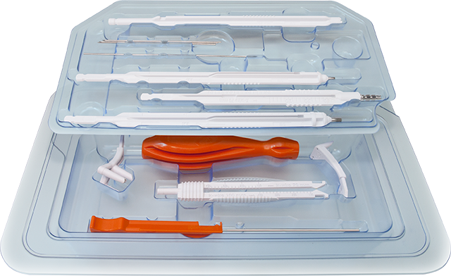
A skin incision is made over the dorsum of the lesser metatarsal phalangeal joint. The distal portion of the metatarsal head is resected at the appropriate level for the existing deformity or disease. Using the supplied Reamer, prepare the medullary canal of the metatarsal. The three laser mark lines refer to the three proximal Implant stem lengths. Refer to Sizing Chart for proximal stem lengths.
JOINT PREPARATION (Phalanx)
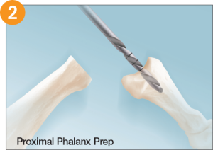
The base of the proximal phalanx is preserved if possible. Using the supplied Reamer, the medullary canal of the proximal phalanx is prepared. Refer to Sizing Chart dimension for distal stem lengths.
IMPLANT SIZING
Place the Implant Trials into the prepared joint in order to select the correct size of Implant. With the Trial in place, load the foot to check that there is no jamming of the Implant. The Trial is then removed and the joint is thoroughly irrigated. The color of the chosen Trial corresponds to the color code on the associated Implant package.
IMPLANT PLACEMENT
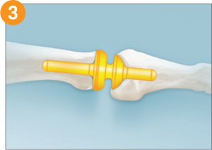
The appropriate Implant is then inserted. Important – HANDS ONLY INSERTION: Do not use forceps or other instruments to grab, grasp and/or insert the Implant. This can damage the Silicone Implant. Excessive handling of the Implant should be avoided. After Implant placement, flush the joint with copious irrigation.

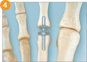
CLOSURERepair and suture the joint capsule, being certain to completely cover the Implant. (Optional technique procedure: Tack down the extensor digitorum longus tendon proximal to the MTP joint, using one or two absorbable sutures, with the digit just slightly plantar flexed at the MTP joint. This will prevent retrograde buckling (dorsal contracture) of the joint while the foot is elevated post operatively, thus allowing the joint capsule to heal in its normal position. The sutures will dissolve after a few weeks, and the tendon and joint will resume function.) Wound closure is performed with sutures of the surgeon’s choice.

Related Products

Quick Contact

We welcome your questions & feedback.